Publications

Influence of vaginal birth on lumbopelvic muscle mechanical properties on urinary incontinence
Authors: Maria Teresa Garzon-Alfaro 1, Ines Cruz-Medel 1, Sandra Alcaraz-Clariana 1, Lourdes Garcia-Luque 1, Maria Cristina Carmona-Perez 1, Juan Luis Garrido-Castro 2, 3, Francisco Alburquerque-Sendin 1, 3, Daiana Priscila Rodrigues-de-Souza 1, 3
Affiliations:
- Department of Nursing, Pharmacology and Physical Therapy, Faculty of Medicine and Nursing, University of Cordoba, Cordoba, Spain
- Department of Computer Science and Numerical Analysis, Rabanales Campus, University of Cordoba, Cordoba, Spain
- 3GC05 Systemic and chronic inflammatory autoimmune diseases of the locomotor system and connectivetissue, Maimonides Biomedical Research Institute of Cordoba (IMIBIC), Cordoba, Spain
Journal: Clinical Rehabilitation - January 2024, Volume 38, Issue 4, Pages 558-568 (DOI: 10.1177/02692155231224058)
-
Field & Applications:
- Medical
- Women's health
- Musculoskeletal disorder
- Validity
- Assessing the muscle mechanical properties of the lumbopelvic region is relevant to improving the management of PF disorders in clinical settings.
Objective: To identify differences in the muscle mechanical properties of the pelvic floor (PF) and lumbar paravertebral (LP) muscles between young nulliparous and uni/multiparous women. Secondarily, specific behaviors, depending on the presence or absence or urinary incontinence (UI), were also researched.
Design: Case-control study.
Setting: Higher education institution.
Participants: One hundred young women participated, divided into two groups depending on whether they had vaginal birth (nulliparous or uni/multiparous). Each group included women with and without UI.
Main measures: A muscle mechanical properties (tone, stiffness, decrement – inverse of elasticity -, and viscoelastic properties: relaxation and creep) assessment of the PF and LP muscles were performed with a hand-held tonometer.
Results: Tone and stiffness of both sides of the PF presented group by UI interaction (p < 0.05), with uni/multiparous women with UI showing higher tone and stiffness compared to multiparous women without UI. In LP muscles, uni/multiparous women showed greater tone and stiffness on the right and left sides [−2.57Hz (95% confidence interval −4.42,−0.72) and −79.74N/m (−143.52,−15.97); −2.20Hz (−3.82,−0.58) and −81.30N/m (−140.66-,21.95), respectively], as well as a decrease in viscoelastic properties compared to nulliparous women [relaxation: 2.88ms (0.31,5.44); creep: 0.15 (0.01,0.30); relaxation: 2.69ms (0.13,5.25); creep: 0.14 (0,0.28), respectively].
Conclusions: Vaginal birth and UI have a differential influence on the muscle mechanical properties of the PF and LP muscles. The determination of muscle mechanical properties by externally applied hand-held tonometry improves the knowledge of the lumbopelvic status, with applicability in clinical and research fields.

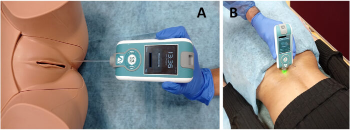
Figure 1. Assessment of the MMPs with MyotonPRO (a) on the left side at PF level (anatomical model) and (b) on the right side at LP level (in vivo). MMPs: mechanical muscle properties; LP: lumbar paravertebral.
Keywords: pelvic floor disorders, patient outcome assessment, women’s health, parity
To summarize, this study showed that different factors are involved in the status of lumbopelvic MMP. Thus, vaginal birth and the presence of UI influence the MMP of the PF and LP muscles. Whereas the PF of uni/multiparous women with UI exhibits higher tone and stiffness than continent women, with no relevant differences compared to nulliparous women with or without UI, the LP muscles of uni/multiparous women show more tone and stiffness and lower viscoelastic properties than nulliparous women, regardless of whether they suffer from UI. Therefore, determining the MMP by externally applied hand-held tonometry is a valid and harmless method for determining MMP, that improves the knowledge of the lumbopelvic status after vaginal birth in women with and without UI, with applicability in clinical and research fields.


