


Publications

Myotonometry Assessment in Children and Adolescents with Pectus Excavatum Included in a Physical Exercise Program
Authors: Marius Zoltan Rezumes 1, Liliana Catan 2, 3, Elena Constanta Amaricai 2, 3, Ada Maria Codreanu 1, 4, Andreea Ancuta Vataman 1, 3, Vlad Laurentiu David 5
Affiliations:
- Doctoral School, Victor Babes University of Medicine and Pharmacy, 300041 Timisoara, Romania
- Research Center for Assessment of Human Motion, Functionality and Disability, Victor Babes University of Medicine and Pharmacy, 300041 Timisoara, Romania
- Department of Rehabilitation, Physical Medicine and Rheumatology, Faculty of Medicine, Victor Babes University of Medicine and Pharmacy, 300041 Timisoara, Romania
- Department of Medicine, Faculty of Medicine, Vasile Goldis Western University, 310038 Arad, Romania
- Department of Pediatric Surgery, Victor Babes University of Medicine and Pharmacy, 300041 Timisoara, Romania
Journal: Healthcare - February 2026, Volume 14, Issue 5, Article no. 613 (DOI: 10.3390/healthcare14050613)
-
Field & Applications:
- Medical
- Treatment evaluation
- Pediatrics
- Orthopedics
- Physiotherapy
- Musculoskeletal disorder
- Musculoskeletal rehabilitation
- Balance / Postural control
- Muscle symmetry
Context/Objectives: Pectus excavatum (PE), the most common anterior chest wall deformity in children and adolescents, impacts posture and is frequently associated with axial deviations due to biomechanical alterations of the spine and the properties of the involved musculature.
Methods: We assessed 35 patients with PE with a Haller index below 3.25, aged between 5 and 17 years, who completed a three months specialized physical exercise program after proper training and instruction by a specialist. All patients were assessed before starting the exercise program and at the end of the treatment. The assessment method used was myotonometry, employing the MyotonPRO device, targeting the trapezius muscle with all three fascicles and the pectoralis major muscle both on the left and the right side, measuring: frequency (Hz), stiffness (N/m), decrement, relaxation time (ms), and the ratio between relaxation time and deformation time (creep).
Results: The analysis of myotonometric parameters reveals a pattern of selective adaptation, predominantly involving the left hemibody in most of the groups analyzed, without significant functional imbalances. This asymmetry may reflect either the functional predominance of the left hemibody during participants’ daily activities or increased activation induced by the exercise program; however, by the end of the intervention, bilateral stability was observed in most parameters.
Conclusions: A three-month physical exercise program in children and adolescents with PE results in improvements in muscle properties, particularly in the pectoralis major and middle trapezius muscles bilaterally, and contributes to the restoration of functional symmetry, thereby supporting the effectiveness of the exercise program in optimizing neuromuscular control, tissue elasticity, and scapular stability.
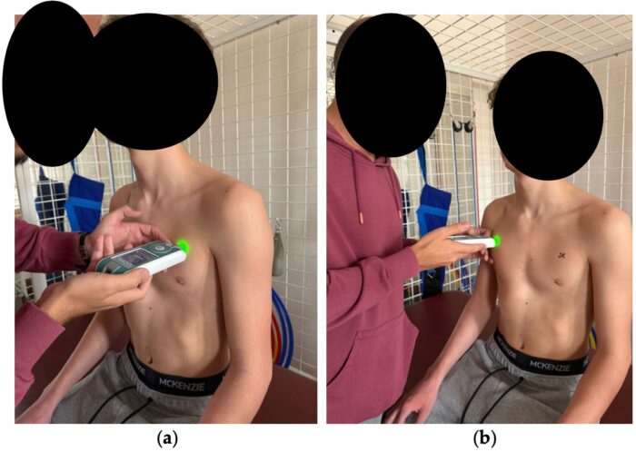
Figure 6. Myotonometric measurement of the pectoralis muscle (a) left (b) right.
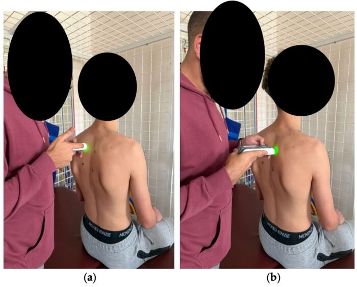
Figure 8. Myotonometric measurement of the middle trapezius muscle (a) left (b) right.
Keywords: pectus excavatum, physical therapy modalities, exercise therapy, muscle tonus, biomechanical phenomena, chest wall, posture, myotonometry, scapular stability, muscle stiffness
The implementation of a 3-month exercise program in children and adolescents with PE leads to improvements in muscle properties, especially in the pectoralis major and middle trapezius muscles bilaterally, and plays a role in restoring functional symmetry, thus supporting the effectiveness of the exercise program in optimizing neuromuscular control, tissue elasticity, and scapular stability.